


Renal Physiology
Kidneys
Physiological anatomy of the kidneys
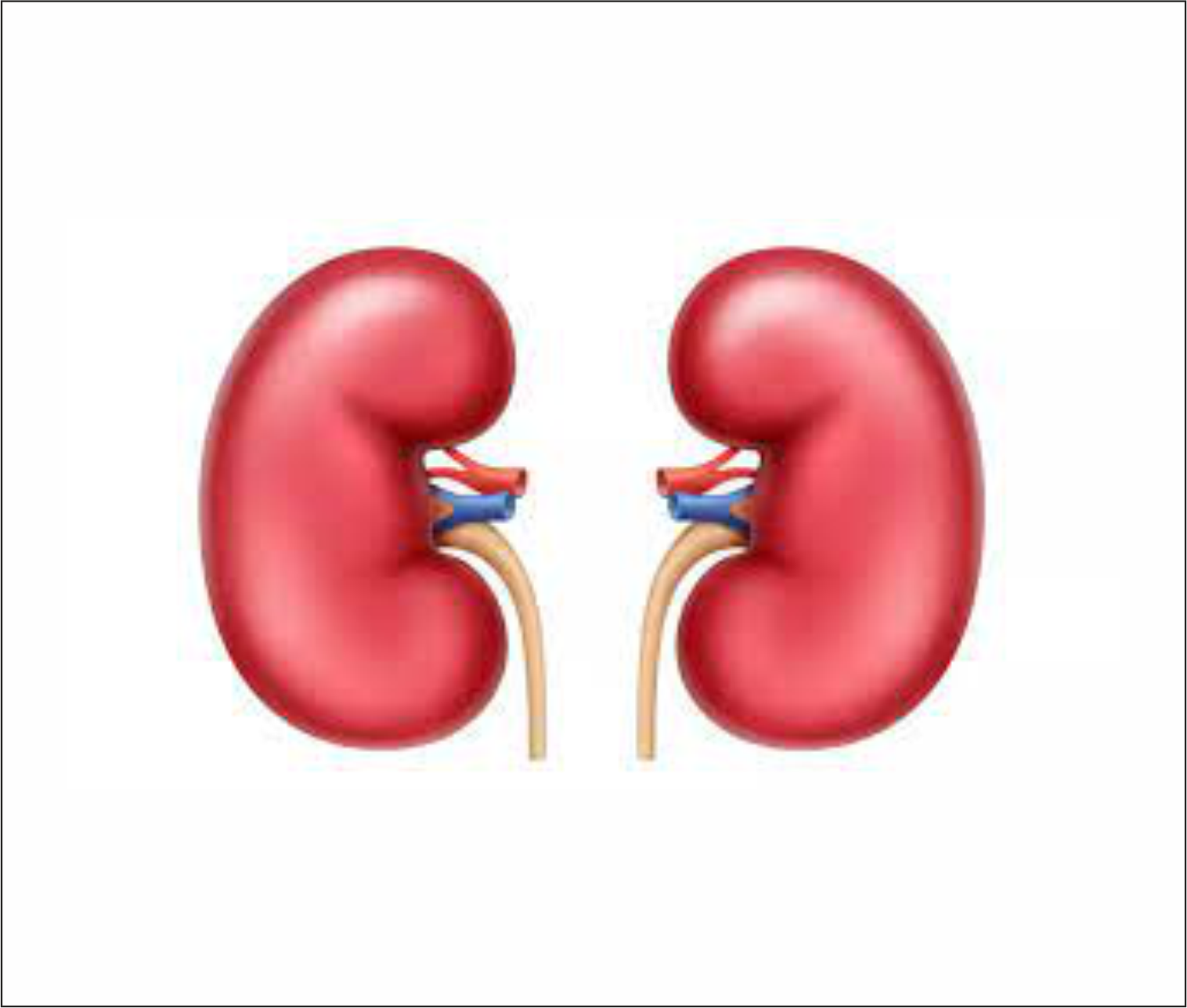
प्रत्येक kidney, abdomen में इसकी posterior abdominal wall पर स्थित रहती हैं। ध्यान रहे, यह peritoneal cavity के भीतर नहीं होतीं बल्कि इसके बाहर retroperitoneal space में स्थित रहती हैं। इनकी केवल anterior surface ही peritoneum के द्वारा covered रहती है। प्रत्येक kidney एक tough fascia से covered रहती है जिसे renal capsule कहते हैं।
प्रत्येक kidney एक bean (राजमा के दाने) के आकार की होती है। इसके बीच का भीतर की ओर दबा हुआ भाग hilum कहलाता है (Latin hilus = opening) ।Kidneys में आने-जाने वाली blood vessels, lymphatics, nerves एवं ureter इसी से होकर गुजरते हैं।
किसी kidney को इसके coronal section में देखने पर इसके दो प्रमुख भाग दिखलाई पड़ते हैं, 1) outer cortex एवं 2) innner medulla ।
Kidneys के functions को समझने के लिए हमें इनके दो प्रमुख structures को समझना होगा। प्रथम nephrons एवं द्वितीय इनकी blood supply
The nephron
Nephrons, kidneys की structural एवं functional units हैं। प्रत्येक kidney में लगभग 10 लाख (1 million) nephrons होते हैं। Kidneys के द्वारा किये जाने वाले समस्त excretory functions इन्हीं nephrons के द्वारा ही सम्पादित होते हैं। इसीलिए इन्हें kidneys की functional unit कहा जाता है। प्रारम्भ में इनमें से प्रत्येक urine formation में सक्षम होता है परन्तु उम्र के साथ-साथ functional nephrons की संख्या में क्रमशः कमी आती जाती है। एक अनुमान के अनुसार, 40 वर्ष के बाद उम्र के प्रत्येक दशक में functional nephrons की संख्या में लगभग 10% की कमी आती जाती है। दूसरे शब्दों में प्रत्येक वर्ष लगभग 1% functional nephrons कम हो जाते हैं। 80 वर्ष की आयु तक functional nephrons की संख्या लगभग 60% ही शेष रहती है। तुम जानते हो कि human body के अधिकाँश organs में उम्र अथवा किसी अन्य प्रकार से होने वाली क्षति से उबरने के लिए अनेकों organs में पर्याप्त reserve उपलब्ध रहता है। इसके अतिरिक्त अनेक tissues (जैसे liver) में regeneration की क्षमता भी आश्चर्यजनक रूप से विद्यमान होती है। परन्तु hepatocytes के विपरीत, nephrons में regeneration की क्षमता नगण्य होती है। इस लिए, उम्र के साथ जैसे-जैसे कुछ nephrons की activity घटती जाती है, अन्य nephrons size में बढ़कर एवं अधिक कार्य करके शरीर का काम चला लेते हैं।
प्रत्येक nephron, दो प्रमुख भागों से मिलकर बनता है, 1) proximal small globular part जिसे Bowman capsule, एवं 2) distal long tubular part ।
Bowman capsule - यह nephron का आरंभिक भाग है जो एक hollow cup के रूप में फूला हुआ होता है। इसीमें blood के filtration का कार्य होता है। Filtration के लिए इसमें से होकर गुजरने वाली blood vessels विभाजित होकर एक capillary network में बदल जाती हैं जिसे glomerulus कहते हैं। Glomerulus - इसमें blood को लाने वाली blood vessel, afferent arteriole कहलाती है (Latin afferrre = लाने वाली) एवं इसमें से blood को ले जाने वाली blood vessel, efferent arteriole कहलाती है (Latin efferre = ले जाने वाली) । Glomerulus से गुजरते समय blood का जो भाग filter होकर Bowman capsule में प्रवेश करता है वह glomerular filtrate कहलाता है।
Tubule - Evolution के साथ-साथ human kidneys में जिस विशिष्ट प्रक्रिया का आरम्भ हुआ वह थी glomerular filtrate की extensive processing । सम्पूर्ण glomerular filtrate को शरीर के बाहर excrete out नहीं कर दिया जाता बल्कि long tubules से होकर निकलते समय इसका भली-भांति निरीक्षण किया जाता है। इस प्रकार, यदि इस filtrate में कुछ ऐसे लाभदायक तत्व भी filter हो गए हों जो शरीर के लिए उपयोगी हों तब उन्हें वापस reabsorb कर लिया जाता है जबकि यदि शरीर के लिए हानिकारक अथवा अवांछित तत्व filter न हो सके हों तब उनको forcefully secrete करा दिया जाता है। Tubules के विभिन्न भागों में यह भिन्न-भिन्न प्रकार के कार्य होते हैं जिसके कारण ही tubules की लम्बाई काफी बढ़ जाती है। इन tubules का अधिकाँश भाग तो अत्यंत घुमावदार सा रहता है जिसे convoluted tubule कहते हैं (convolution = लहरदार अथवा घुमावदार) । Tubules के मध्य का भाग straight एवं किसी hair pin की भांति होता है जिसे loop of Henle कहते हैं। इस प्रकार यह loop of Henle, किसी tubule को दो भागों में विभाजित करता है, 1) proximal convoluted tubule (PCT) एवं 2) distal convoluted tubule (DCT)।
Collecting tubule - प्रत्येक nephron, एक connecting tubule के माध्यम से एक collecting tubule में drain करता है जो पुनः collecting duct में drain करती है।
Kidney में यह nephrons किसी कलियों के गुच्छे की भाँति व्यवस्थित रहते हैं। जिस प्रकार किसी गुच्छे में कलियों वाला ऊपरी भाग फूला हुआ होता है एवं डंठल वाला निचला भाग अपेक्षाकृत पतला होता है, उसी प्रकार, nephrons का यह गुच्छा भी Bowman capsule एवं convoluted tubules के कारण ऊपर से चौड़ा एवं पतले-पतले loop of Henle, collecting tubules एवं collecting ducts के कारण नीचे से पतला होता है। Cone अथवा pyramid के आकार के होने के कारण ही इन गुच्छों के निचले भाग को renal pyramid कहते हैं। प्रत्येक kidney में ऐसे 8-10 pyramids होते हैं।
अब हम पुनः kidneys के coronal section पर लौटते हैं। सभी nephrons के glomeruli (pleural of glomerulus) एवं PCT, renal cortex में स्थित होते हैं। PCT से निकलने वाला loop of Henle, renal cortex से निकलकर, renal medulla में लूप बनता हुआ पुनः renal cortex में लौट आता है। Renal cortex से नीचे उतर कर loop of Henle के medulla की ओर जाने वाले भाग को इसकी descending limb एवं वहां से ऊपर चढ़ते हुए cortex में वापस लौटने वाले भाग को इसकी ascending limb कहते हैं। इस प्रकार DCT पुनः renal cortex में ही मिलती है। यहीं यह DCT, connecting tubules के माध्यम से collecting tubules में खुलती है। क्योंकि यह प्रारंभिक collecting tubules, renal cortex में स्थित होती हैं अतः इन्हें cortical collecting tubules कहते हैं।
Cortical and medullary collecting duct - Individual nephron से drain होती हुई इस urine को kidney से बाहर ले जाने के लिए cortical collecting tubules को पुनः renal medulla की ओर मुड़ना पड़ता है। Renal medulla की ओर बढ़ते हुए यह cortical collecting tubules परस्पर मिलती जाती हैं। 8-10 cortical collecting tubules के मिलने से एक larger cortical collecting duct का निर्माण होता है। Renal medulla में पहुँचकर यह medullary collecting duct कहलाती है। अनेकों medullary collecting ducts के मिलने से इन collecting ducts का diameter क्रमशः बढ़ता जाता है। यह larger collecting ducts अंततः renal pyramid की conical tip से निकलती हैं। इन tips को renal papilla (pleural - papillae) कहते हैं। प्रत्येक kidney में लगभग 250 very large collecting ducts होती हैं जिनमें से प्रत्येक लगभग 4000 nephrons को drain करती है।
Urinary collecting system - प्रत्येक renal papilla एक cup के आकार के minor calyx में खुलता है (Latin kalux = cover)। यह उसी प्रकार हुआ जैसे किसी फूलों के गुच्छे को किसी चौड़े मुंह के बर्तन में रख दिया जाये। इस प्रकार renal papillae से निकलने वाली सभी collecting ducts, एक minor calyx में drain करती हैं। दो या तीन minor calyces (pleural of calyx) के मिलने से एक major calyx बनती है। पुनः दो या तीन major calyces मिलकर renal pelvis में drain करती हैं। यह renal pelvis, kidney के hilum से बाहर निकलकर ureter में drain करती है।
आओ nephron से लेकर collecting system के क्रम को एक बार पुनः समझ लेते हैं।
Bowman capsule - PCT - loop of Henle - DCT - connecting tubule - cortical collecting tubule - cortical collecting duct - medullary collecting duct - larger collecting duct - minor calyx - major calyx - pelvis - ureter
आओ kidney के किस भाग में nephrons के क्या-क्या भाग मिलते हैं इनको भी समझ लेते हैं।
Renal cortex = Bowman capsules with glomeruli and blood vessels + PCT + DCT + connecting tubule + cortical collecting tubules and ducts
Renal medulla = Loop of Henle + medullary collecting ducts + very large collecting ducts + minor and major calyces + pelvis + medullary interstitial tissue
Dimensions of a nephron
Glomerulus - ~200 micrometer in diameter
Tubules - ~55 micrometer in diameter, 45-65 mm in length
PCT - ~15 mm
DCT - ~5 mm
Collecting duct - ~20 mm
Renal vascular system
Renal artery and its branches - प्रत्येक kidney जिस artery से अपनी blood supply प्राप्त करती है उसे renal artery कहते हैं। यह descending aorta के abdominal part से निकलकर hilum से kidney में प्रवेश करती है।
Kidney में प्रवेश करने के पश्चात् renal artery 2 या 3 segmental srteries में विभाजित हो जाती है।
प्रत्येक segmantal artery पुनः 2 या 3 interlobar arteries में विभाजित हो जाती है। अपने नाम के अनुसार यह interlobar arteries, दो lobes या दो renal pyramids के मध्य चलते हुए renal cortex की ओर बढ़ती हैं।
Cortex में पहुंचकर यह interlobar artery, renal pyramid की outer surface पर एक arc की भांति घूम जाती है। इसीलिए अब इसे arcuate artery कहते हैं।
इस arcuate artery से अनेकों smaller perpendicular arteries निकलती हैं जो renal cortex की outer surface की ओर बढ़ती हैं। इन्हें interlobular arteries कहते हैं।
Glomerular capillary network - इन्हीं interlobular arteries से अनेकों very small afferent arterioles निकलती हैं जो glomerulus को blood supply करती हैं। इन्हीं की branching से glomerular capillary network का निर्माण होता है।
Glomerular capilaries के fusion से पुनः एक arterioles का निर्माण होता है जिसे efferent arteriole कहते हैं। यहाँ ध्यान दो, anatomy के सामान्य सिद्धांत के अनुसार capillaries से निकलने वाली vessel को vein कहा जाता है परन्तु glomerulus से निकलने वाली vessel को arteriole ही कहा गया है। वास्तव में ऐसा इसलिए क्योंकि यह efferent arteriole, पुनः एक अन्य capillary network को blood supply करती है (एवं इस प्रकार से एक artery के रूप में ही व्यवहार करती है)।
Peritubular capillary network - यह efferent arteriole, loop of Henle के descending limb की ओर बढ़ते हुए पुनः विभाजित होकर एक दूसरे peritubular capillary network का निर्माण करती है जो loop of Henle के चारों ओर रहता है। इस प्रकार से renal circulation में एक vascular portal system देखने को मिलता है जिसमें एक capillary circulation से निकलने वाली blood vessels पुनः एक अन्य capillary circulation को उत्पन्न करती हैं।
Veins - Loop of Henle के ascending limb पर यह capillaries परस्पर मिलकर एक vein का निर्माण करती हैं जो respective arteries के ही साथ-साथ चलती हुई interlobular vein, arcuate vein, interlobar vein एवं segmental vein से होते हुए renal vein के माध्यम से inferior vena cava में blood को drain करती हैं।
Renal circulation
शरीर के अन्य organs की अपेक्षा renal circulation की कुछ अपनी विशिष्टताएं हैं।
Renal blood flow is much much more than it’s requirements - दोनों kidneys का वजन, शरीर के कुल वजन का केवल 0.4% होता है। इस पर भी total cardiac output का लगभग 22% भाग (1100 ml/min), दोनों kidneys को पहुँचता है। इसके दो प्रमुख कारण हैं।
Excretion सम्बंधित गतिविधियों के लगातार चलने के कारण kidneys की अपनी metabolic demand, अन्य organs की अपेक्षा अधिक होती है। परन्तु यह इतनी भी अधिक नहीं होती जिसके लिए इतनी अधिक blood supply की आवश्यकता हो।
वास्तव में, kidneys को इतनी अधिक मात्रा में blood की supply, शरीर को अपनी आवश्यकता (metabolic waste products के excretion) के कारण करनी पड़ती है। इसी high blood flow के माध्यम से ही शरीर, glomerulus से पर्याप्त मात्रा में filtration करवा पाता है जिससे metabolic waste products को excrete out करवाया जा सके एवं water एवं electrolyte concentration को नियंत्रण में रखा जा सके।
Sodium reabsorption through Na-K ATPase pump is the reason for the higher metabolic demand of the kidneys - Blood supply के साथ-साथ kidneys को oxygen भी प्रचुर मात्रा में मिलती है। Weight to weight basis पर kidneys की oxygen demand brain की भी अपेक्षा दो गुनी होती है। यहाँ प्रश्न यह उठता है कि kidneys में ऐसी कौन सी metabolic activity चल रही है जिसके कारण इसकी metabolic demand इतनी अधिक है? याद करो, शरीर की basal metabolic demand का एक बड़ा भाग Na-K ATPase pump को चलाने में ही व्यय हो जाता है। Glomerulus से जितनी अधिक मात्रा में filtration होता है, Na+ की भी उतनी ही अधिक मात्रा उसके साथ filter होती जाती है। Tubular cells के द्वारा इसी Na+ को reabsorb करने में, Na-K ATPase pump बड़ी भूमिका निभाता है। यदि glomerular filtration अधिक बढ़ता है तब उसके साथ-साथ Na filtration भी बढ़ता है एवं अधिक Na को reabsorb कराने के लिए Na-K ATPase pump के माध्यम से kidneys की metabolic demand भी उतनी ही अधिक बढ़ती जाती है।
Renal blood flow is non-homogenously distributed within kidneys - इतनी अधिक मात्रा में blood supply होने पर भी kidney के सभी भागों को समान मात्रा में blood नहीं मिल पाता। Total blood supply का लगभग 98% भाग तो renal cortex में glomerulus से filtration एवं tubules से reabsorption की प्रक्रिया में ही प्रयुक्त होता हुआ, renal veins में वापस लौट जाता है। केवल 1-2% भाग ही vasa recta के माध्यम से renal medula को प्राप्त हो पाता है। इसके कारण, renal medulla (विशेषतयः medulla tip या renal papilla पर) में ischemic damage की सम्भावना हमेशा बनी रहती है।
Renal blood flow can be autoregulated - शरीर के अधिकाँश organs, अपनी nutrients एवं oxygen demands के अनुसार, अपने blood flow को कुछ मात्रा में autoregulate कर सकते हैं। क्योंकि, kidneys में blood flow पहले से ही आवश्यकता से काफी अधिक होता है अतः kidneys को अपने लिए इस autoregulation की आवश्यकता नहीं पड़ती। Kidneys अपने blood flow का autoregulation, इसके द्वारा हो रहे glomerular filtration (GFR) एवं water एवं electrolytes के excretion को नियंत्रित करने के लिए करती हैं। इसके द्वारा kidneys, mean arterial pressure के 70 mm Hg तक घट जाने अथवा 170 mm Hg तक बढ़ जाने पर भी, GFR को 10% से भी कम घटने अथवा बढ़ने से रोकने में सफल हो जाती हैं। Kidneys द्वारा renal blood flow का यह autoregulation दो प्रकार से संपन्न हो सकता है।
Intrinsic feedback mechanism - जो isolated blood perfused kidneys में भी कार्य करता है, एवं
Myogenic mechanism - जिसमें BP के बढ़ने पर afferent arteriole constrict होकर glomerular perfusion घटा देती है तथा BP का घटने पर यह dilate होकर glomerular perfusion को बढ़ा देती है।
Cortical and juxtaglomerular nephrons
जिस प्रकार evolution से साथ-साथ nephrons में tubular functions (reabsorption एवं secretion) का विकास हुआ, ठीक उसी प्रकार terestrial life में पानी की कमी होने की सम्भावना से बचाव के लिए urinary concentrating ability का भी विकास हुआ। इस कार्य के संपादन हेतु nephrons की एक नयी प्रजाति उत्पन्न हुई जो अन्य nephrons से अलग, water एवं salt conservation के लिए विशेष रूप से बनी थी। जहाँ kidneys के लगभग 85% nephrons renal cortex में ही मिलते हैं एवं उनके loof of Henle का केवल एक छोटा सा हिस्सा ही medulla के बाहरी भागों तक पहुंच पाता है वहीँ लगभग 15% neurons, renal cortex एवं medulla के junction पर ही स्थित होते हैं। Medulla के समीप होने के कारण ही इन्हें juxta-medullary nephrons (juxta = निकट) कहा जाता है। इन nephrons की कुछ अत्यंत महत्वपूर्ण विशेषतायें निम्नांकित हैं -
Long in length - इनका loop of Henle, cortical nephrons की अपेक्षाकृत काफी अधिक लम्बा होता है जो medulla में काफी नीचे तक उतरता चला जाता है।
इनके loop of Henle की cells भी cortical nephrons की भांति एक समान न होकर दो भिन्न-भिन्न प्रकार की होती हैं, highly permeable thin cells एवं less permeable thick cells । thin Thin and thick segments - Descending limb तो लगभग पूरा ही thin cells के द्वारा बना होता है जबकि ascending limb में thin एवं thick दोनों प्रकार के cells मिलते हैं। इस प्रकार, loop of Henle का descending limb, thin segment का बना होता है जबकि ascending limb का proximal part, thin segmant एवं distal part, thick segment का बना होता है।
Presence of vasa recta - इनकी peritubular capillary network में tortuous (घुमावदार) capillaries के अतिरिक्त एक विशिष्ट straight vessel मिलती है जिसे vasa recta कहते हैं (vasa = vessel; recta = straight) । वास्तव में, glomerulus से निकलने वाली efferent arteriole ही loop of Henle के ascending limb के साथ-साथ (parallel) नीचे उतरती है एवं पुनः descending limb के साथ-साथ कुछ दूर तक ऊपर चढ़ती है। इसके बाद यही vasa recta, tortuous capillary network में विभाजित हो जाती है। इस प्रकार, loop of Henle का thin segment, vasa recta के साथ एवं thick segment, peritubular capillary network के साथ रहता है। ध्यान रहे, loop of Henle में glomerular filtrate का flow, descending limb से ascending limb की ओर होता है जबकि vasa recta में blood flow, descending limb से ascending limb की ओर होता है। दो parrelal tubes में परस्पर विपरीत दिशाओं में हो रहे इस flow को counter current system कहते हैं (counter = विपरीत; current = धारा)। Loop of Henle के permeable thin segment के साथ-साथ विपरीत दिशा में होने वाला यह blood flow, tubular fluid की concentration को महत्वपूर्ण रूप से प्रभावित करता है जिसके विषय में हम बाद में पढ़ेंगे।
इस प्रकार, juxtaglomerular nephrons का अधिकाँश भाग तो vasa recta के साथ रहता है एवं केवल ascending limb का distal thick segment ही peritubular capillary network के साथ रहता है। इसी कारण से juxtaglomerular nephrons का peritubular capillary network, cortical nephrons की तुलना में काफी छोटा होता है।
Functions of the kidneys
Kidney के functions के विषय में पूछने पर संभव है कि तुम्हारे मन में केवल एक ही उत्तर आये, metabolic waste products का excretion । परन्तु ध्यान रहे, kidney का function केवल excretion मात्र ही नहीं है। इसका एक अन्य अत्यंत महत्वपूर्ण कार्य है, शरीर में water एवं electrolyte balance को बनाये रखना जिससे शरीर के internal milieu को समान बनाये रखा जा सके। आओ इसके सभी कार्यों पर एक दृष्टि डालते हैं।
Excretion of metabolic waste products and foreign materials - शरीर में विभिन्न metabolic processes द्वारा अनेक प्रकार के waste products उत्पन्न होते हैं जैसे urea (amino acids से), creatinine (muscle creatine से), uric acid (nucleic acids से) एवं bilirubin (hemoglobin से)। इन सबका excretion kidneys के ही माध्यम से होता है। यही kidneys का सर्वप्रमुख कार्य है। इनके अतिरिक्त, drugs एवं इनके metabolites तथा शरीर में विभिन्न मार्गों से आये हुए chemicals एवं toxins भी kidneys के द्वारा ही excrete out किये जाते हैं।
Regulation of water and electrolyte balance - Water intake के 1 L से भी कम या 20 L से भी अधिक हो जाने पर भी kidneys water का excretion घटा एवं बढ़ाकर, total body water level में विशेष प्रभाव नहीं पड़ने देतीं। इसी प्रकार, salt intake के भी 10 गुना अधिक बढ़ जाने पर भी यह sodium excretion बढ़ाकर, body fluids में sodium concentration को normal limits में ही बनाये रखने में भी मददकारी होती हैं।
Regulation of osmolality of body fluids - Water एवं electrolyte concentration को बनाये रखने से ही body fluids की osmolality भी normal limits में रखने में मदद मिलती है।
Regulation of acid base balance - H+ एवं HCO3- के excretion को प्रभावित करके kidneys शरीर में acid base balance बनाये रखने में भी सहायक होती हैं। उपरोक्त सभी प्रक्रियाओं की चर्चा हम आगे करेंगें।
Regulation of blood pressure - Kidneys, blood pressure के short term एवं long term control, दोनों में महत्वपूर्ण भूमिका निभाती हैं। BP के घटने से kidneys की juxtaglomerular cells, renin उत्पन्न करती हैं। Liver में यह renin, angiotensinogen को angiotensin I में बदल देता है जो पुनः angiotensin converting enzyme की मदद से angiotensin II में बदला जाता है। यह angiotensin II एक potent vasoconstrictor agent है जो vasoconstriction कराकर BP को बढ़ने में मदद करता है। Renin synthesis के अतिरिक्त, angiotensin I से angiotensin II का निर्माण भी kidneys में ही होता है।
Activation of vitamin D - Skin में UV rays के प्रभाव में cholesterol से vitamin D (cholecalciferol) का निर्माण होता है जो liver के द्वारा 25 hydroxy vitamin D (calcidiol) में परिवर्तित कर दिया जाता है। परन्तु vitamin D की यह दोनों ही forms active नहीं होतीं। इनके activation में kidneys अत्यंत महत्वपूर्ण भूमिका निभाती हैं। Kidneys में parathyroid hormone की मदद से इस 25-hydroxy vitamin D के 1-alpha hydroxylation से 1,25-dihydroxy vitamin D का निर्माण होता है जो calcium एवं phosphorus की metabolism में अत्यंत महत्वपूर्ण भूमिका निभाता है। Severe kidney disease में vitamin D की इस active form की कमी से metabolic bone diseases (जैसे osteomalacia एवं osteoporosis) उत्पन्न होने लगती हैं।
Production of erythropoietin - Kidneys में erythropoietin का भी निर्माण होता है जो bone marrow में hematopoietic stem cells को stimulate करके उनसे erythrocytes का निर्माण करवाती है। Erythropoietin की कमी, severe kidney disease में anemia का एक प्रमुख कारण होती है।
Production of glucose by gluconeogenesis - ध्यान रहे, overnight fasting के पश्चात् जब केवल glycogenolysis के द्वारा blood glucose level को बनाये रखना संभव नहीं हो पाता तब liver में gluconeogenesis की प्रक्रिया से glucose का निर्माण आरम्भ होता है। Prolonged fasting के बाद यह कार्य अकेले liver के द्वारा भी संभव नहीं हो पाता। ऐसे में kidneys भी इसमें अपना महत्वपूर्ण योगदान देती हैं। वास्तव में, fasting के prolonged होने के साथ-साथ gluconeogenesis में liver की अपेक्षा kidneys का योगदान बढ़ता जाता है। याद रहे, kidneys द्वारा हो रही इस gluconeogenesis में मुख्यतः amino acid glutamine का प्रयोग होता है। Severe kidney disease में gluconeogenesis की प्रक्रिया में कमी आने से hypoglycemia की सम्भावना बढ़ जाती हैं।
Formation of urine by the kidneys
तुम जानते हो कि kidneys का प्रमुख कार्य है शरीर में उपस्थित metabolic waste products का urine के रूप में excretion । Urine formation की सम्पूर्ण प्रक्रिया मुख्यतः तीन चरणों में संपन्न होती है।
Filtration - सर्वप्रथम, glomerular capillary network से गुजरते समय, blood में उपस्थित अनेक metabolic waste products (एवं इनके साथ-साथ शरीर के अनेकों useful constituents), filter होकर Bowman capsule में आ जाते हैं। इस filtered material को glomerular filtrate कहते हैं।
Reabsorption - Bowman capsule से निकलकर यह glomerular filtrate, tubule में पहुँचता है। Nephrons में इस लम्बे tubular flow के दौरान शरीर का यह प्रयास रहता है कि glomerulus से अनावश्यक रूप से filter हुए useful constituents के loss को रोकने के लिए उन्हें वापस reabsorb करा लिया जाये।
Secretion - इसके विपरीत, शरीर का पुनः यह प्रयास रहता है कि कुछ metabolic waste products जो किन्हीं कारणों से glomerulus के द्वारा filter होने से छूट गए हों, उन्हें भी nephrons में इस लम्बे tubular flow के दौरान किसी प्रकार से blood से निकालकर tubular fluid में secrete करा दिया जाए।
Excretion - उपरोक्त तीनों प्रक्रियाओं के संपन्न होने के पश्चात् जो अंतिम fluid, शरीर से बाहर निकाला जाता है वही urine कहलाता है। इस सम्पूर्ण प्रक्रिया को एक equation के रूप में निम्नांकित रूप से लिखा जा सकता है।
Excretion = Filtration - Reabsorption + Secretion
आओ, इन तीनों प्रक्रियाओं को विस्तार से समझते हैं।
Filtration
Glomerular filtration membrane - Filtration की प्रक्रिया को समझने के पूर्व glomerular filtration membrane की संरचना को समझना आवश्यक होगा जिससे होकर यह प्रक्रिया संचालित होनी है। Filtration के दौरान filtrate को तीन structures से होकर गुजरना पड़ता है। 1) Capillary endothelium, 2) glomerular basement membrane, जिसे basal lamina भी कहते हैं, एवं 3) epithelial cells of Bowman capsule ।
Capillary endothelium - शरीर की सभी capillary walls, single cell thick होती हैं जो endothelium से बनती है। परन्तु इस endothelium का arrangement प्रत्येक organ की अपनी आवश्यकतानुसार भिन्न-भिन्न हो सकता है। जिन organs में capillaries से हो रहे fluid movement को रोकने की आवश्यकता होती है वहां की endothelial cells tightly packed होती हैं एवं उनके बीच gap junctions नहीं होते (जैसे brain)। इसके विपरीत, जिन organs में इन capillaries से होकर fluid एवं solutes की बड़ी मात्रा को निकलना होता है (जैसे liver एवं kidneys) वहां की endothelial cells के मध्य large gap junctions होते हैं। Glomerular capillaries में इन gap junctions या pores को fenestrations कहते हैं जो 70-90 nm के diameter तक हो सकते हैं। यह fenestrations, glomerulus से होने वाले fluid एवं solutes के filtration में विशेष रूप से सहायक होते हैं।
Capsular epithelium - Glomerulus अपने चारों ओर से Bowman capsule की epithelium के द्वारा घिरा रहता है। Filtration की प्रक्रिया में मदद करने के लिए यह epithelial cells, capillary endothelium को पूरी तरह से cover नहीं करतीं। इन epithelial cells से निकलने वाले long foot like processes (pseudopodia) ही endothelial cells के संपर्क में आते हैं। इससे इन foot processes के मध्य अनेकों gaps छूट जाते हैं जिन्हें slit pores कहते हैं। यह slit pores भी लगभग 25 nm चौड़े होते हैं जिससे होकर fluid एवं small molecules सरलता से filter हो पाते हैं।
Glomerular basement membrane (basal lamina) - Capillary endothelium एवं capsular epithelium के मध्य connective tissues (मुख्यतः collagen एवं proteoglycans) से बनी एक thin membrane रहती है जिसे glomerular basement membrane (GBM) कहते हैं। 4 nm तक के molecules तो इस GBM से होकर बड़ी सरलता से निकल जाते हैं एवं 4-7 nm के molecules कुछ resistance के साथ। 8 nm से बड़े molecules, GBM से होकर नहीं निकल पाते। यहाँ एक महत्वपूर्ण तथ्य को ध्यान में रखने की आवश्यकता है कि albumin molecule का diameter 7 nm होते हुए भी वह GBM को पार नहीं कर पाता। वास्तव में protein जैसे महत्वपूर्ण molecule को filter होने से बचने के लिए प्रकृति ने इसमें एक सुरक्षा कवच भी दिया है। अपने sialoprotein content के कारण GBM, negatively charged होती है। यही negative charge, negatively charged, anionic albumin molecules को repel करते हुए उन्हें filter होने से रोकता है। ध्यान रहे, glomerular capillary endothelial cells एवं capsular epithelial cells पर भी negatively charged protein molecules लगे होते हैं जो plasma albumin को repel करते हुए उनको filter होने से रोकते हैं।
Adaptations in renal microcirculation for effective filtration
उपरोक्त वर्णन से तुम समझ चुके होंगे कि kidneys की एक अनूठी कार्यप्रणाली है। इसका प्रयास होता है कि fluid एवं इसमें dessolved constituents को पहले जितना अधिक से अधिक filter करा सके वह करा दे एवं इसके उपरान्त क्रमशः अपनी आवश्यकतानुसार useful constituents को reabsorb करा ले एवं harmful waste products को excrete हो जाने दे। इसकी सर्वप्रथम आवश्यकता है, एक प्रभावी filtration mechanism । वास्तव में, glomerulus का विकास ही इस प्रकार से हुआ कि उसमें से होकर गुजरने वाला blood, प्रभावी रूप से filter हो सके। इसके लिए glomerular capillary network में निम्नांकित विशेषताएं होती हैं।
Large fenestrations in glomerular capillary endothelium - जिनसे होकर 70 nm तक के molecules निकल सकते हैं।
Slit pores between foot processes of Bowman capsular epithelium - जिनसे होकर 25 nm तक के molecules निकल सकते हैं।
High hydrostatic pressure in glomerular capillaries - स्वाभाविक रूप से कोई fluid, जितना अधिक pressure किसी filtering membrane पर डालेगा, उसका filtration उतना ही अधिक हो सकेगा। इसके लिए भी glomerulus की afferent arteriole का diameter, efferent arteriole के diameter से अधिक होता है। अतः, glomerulus में blood आता सरलता से है परन्तु बाहर निकलता कुछ resistance के साथ है। इससे glomerular capillaries में hydrostatic pressure (~60 mm Hg), शरीर की अन्य capillaries (~17 mm Hg) की तुलना में अधिक हो जाता है। यह glomerular filtration में मदद करता है।
URINARY BLADDER
आओ micturition reflex को समझते हैं। परन्तु इसके लिये पहले urinary bladder के innervation को समझना आवश्यक है एवं innervation जानने से
पहले urinary bladder के musculature को समझना होगा। इसीलिये हम urinary bladder की muscles से ही आरंभ करते हैं।
ध्यान दो, urinary bladder में दो प्रकार की muscles हैंµ पहली bladder proper या detrusor muscle एवं दूसरी sphincteric muscle। पुनः sphincters भी दो प्रकार के होते हैंµ पहला internal sphincter जो वास्तव में detrusor muscle का ही thickened part है एवं दूसरा external sphincter।
क्या तुम बता सकते हो कि यह सभी muscles एक ही प्रकार की हैं या भिन्न-भिन्न प्रकार की? याद करो, urinary bladder एक visceral organ है तथा इसकी muscle smooth होगी। इस प्रकार detrusor muscle एवं इसका ही thickened part – internal sphincter वास्तव में smooth muscle के बने हैं, जो हमारे voluntary control में नही हैं। इसके विपरीत, external sphincter एक striated या skeletal muscle से बना है, जिसके द्वारा हम micturition process पर voluntary control रख सकते हैं।
इस प्रकार urinary bladder का innervation भी involuntary (अर्थात autonomic) एवं voluntary (अर्थात somatic) nervous system दोनों से होना चाहिये। Autonomic nervous system (ANS) के भी दो भाग होते हैं- sympathetic (SNS) एवं parasympathetic (PNS)। तुम जानते हो कि SNS किसी stressful condition के समय activated रहता है। स्वयं सोचो, stress के समय तुम क्या चाहोगे, urinate करने जाना या उसको रोक कर stress से निबटने का प्रयत्न करना? इस प्रकार SNS के प्रभाव में urination को रोकने के प्रयास सक्रिय रहेंगे, जिसका अर्थ हुआ detrusor muscle का relaxation एवं internal sphincter का contraction। इसके विपरीत, PNS विश्राम या relaxation के समय activated रहता है। अतः urination के लिये PNS को activate होना होगा, जिसके द्वारा detrusor muscle contract करेगी एवं internal sphincter relax। परन्तु urination के इस passive act पर हमारा voluntary control भी है, जो external sphincter के द्वारा सम्पन्न होता है। अतः urination की प्रक्रिया आरंभ करने के लिये voluntarily external sphincter को relax कराना होगा।
Sympathetic fibres, T 10,11,12 spinal segments से निकलकर inferior splanchnic nerve के द्वारा inferior mesenteric ganglion पहुंचते हैं जहां से वह hypogastric nerve के माध्यम से pelvic plexus या inferior hypogastric ganglion एवं वहां से urinary bladder पहुंचते हैं।
Parasympathetic fibres S 2, 3, 4 segments से निकल कर hypogastric nerve के माध्यम से pelvic plexus या inteiror hypogastric ganglion होते हुए urinary bladder तक पहुंचते हैं।
Somatic fibres S 2, 3, 4 segments से निककर internal pudendal nerves के माध्यम से external sphincter तक पहुंचते हैं।
जरा सोचो, urination की आवश्यकता कब होगी? अवश्य ही जब urinary bladder भरा हो। इसका अर्थ हुआ कि urinary bladder के distension के
sensations भी CNS तक पहुंचने चाहिये। जरा सोचो, यह किस nerve द्वारा जाने चाहिये? वास्तव में urinary bladder की filling से उत्पन्न हुआ pressure एवं overfilling से उत्पन्न हुआ pain sensation, दोनों, pudendal nerve के द्वारा ही spinal segments S 2, 3, 4 एवं वहां से higher centres तक ले जाये जाते हैं। इसके अतिरिक्त अन्य स्थानों की smooth muscles की भांति ही bladder के pain sensations sympathetic fibres द्वारा भी ले जाये जाते हैं।
अब सोचो, urination अथवा micturition की प्रक्रिया क्या होगी? Urinary bladder की filling से इसके pressure sensations pudendal nerve द्वारा spinal S 2, 3, 4 segments तक पहुंचते हैं, जहां से वह एक autonomic reflex प्रक्रिया की भांति PNS को उत्तेजित कर S 2,3,4 segments से ही parasympathetic discharge बढ़ा देते हैं, जिससे detrusor muscle contract करे व internal sphincter relax। इस प्रकार urination आरंभ हो जाता है। यदि परिस्थितियाँ अनुकूल हों तब व्यक्ति external sphincter को voluntarily खोलकर urination आरंभ हो जाने देगा अन्यथा इसे बंद ही रख कर इस micturition reflex को रोक देगा।
जरा ध्यान करो, कई बार urinary bladder की overfilling हो जाने पर भी यदि urination की सुविधा उपलब्ध न हो सके तब तुम क्या करते हो? ऐसे में केवल external sphincter के contraction से ही काम नही चल पाता एवं समस्त perineal muscles यहां तक कि anal sphincter तक को contract कराना पड़ जाता है। इस प्रकार यह perineal muscles भी urination को voluntarily inhibit करने में सहायक होती हैं। इसके विपरीत overdistended urinary bladder के साथ urination आरंभ करते समय urinary flow कुछ समय तक रुका-रुका सा लगता है, जिसके लिये abdominal muscles के contraction के द्वारा intrabdominal pressure बढ़ाकर detrusor muscles पर बाहर से जोर लगाना पड़ता है। इस प्रकार यह abdominal muscles भी micturition के flow को बढ़ाने में भी मदद कर सकती हैं।
जरा सोचो, क्या males अथवा females के micturition process में कोई अंतर हो सकता है? इस तथ्य को समझने के लिये तुम्हे दोनों के pelvic structures के anatomical dffierences को समझना होगा। याद करो, female urithera केवल 4 cm लंबी होती है, जबकि male urethera 20 cm। इसके अतिरिक्त male urethra का एक लंबा भाग penis से pass होता है, जहां वह penile muscles एवं दूसरे penile structures द्वारा घिरा रहता है। इस प्रकार तुम स्वयं यह अनुमान लगा सकते हो कि male में penile urethra urination में एक partial hindrance उत्पन्न करता है। Females में जहां urination process आरंभ होने के बाद gravity के प्रभाव में लगातार होता हुआ अंत में स्वयं ही पूर्ण हो जाता है, वहीं males में urination के अंतिम भाग में जब intravesical pressure कम हो जाता है तथा कुछ urine urethra में भी रुकी रहती है, तब इसे abdominal muscles एवं bulbocavernosus muscles के contraction से बाहर निकालने के लिये कुछ extra efforts करने पड़ते हैं।
जरा सोचो, urinary bladder के contract करने पर urine का flow urethra में ही क्यों होता है, वापस ureter में क्यों नही?
यह अनुमान तो तुम स्वयं लगा सकते हो कि urinary bladder के contract करने पर यदि urine वापस ureter में पहुंचने लग जाये तो यह urination की प्रक्रिया को ही effective बना देगा। इसके अतिरिक्त यह backflow किसी प्रकार के lower urinary tract infection को भी kidneys तक पहुंचा सकता है, जिसके गंभीर दुषपरिणाम हो सकते हैं। इसीलिये प्रकृति ने इस backflow को रोकने की व्यवस्था भी की है। वास्तव में दोनों ureters, urinary bladder में खुलने से पहले उसकी wall को 1- 2 cm तक obliquely cross करते हैं। इसका अर्थ हुआ कि bladder की outer wall से enter होने के बाद ureter कुछ दूरी तक bladder wall के अंदर travel करते हैं तथा फिर inner wall से bladder के lumen में enter होते है। Ureter का यह intravesical route एक valve की तरह कार्य करता है। Bladder में हो रही urinary filling intravesical pressure को बढ़ाती है, जिससे ureter की inner wall में opening बाहर की तरफ होती है एवं एक valve की भांति इसे बंद कर देती है। इसके अतिरिक्त ureter का intravesical part भी इस pressure से compress होकर बंद हो जाता है। इसी valve के dysfunction से intravesical pressure बढ़ने पर urine ureter में leak करने लगती है, press जिसे vesico ureteric reflex (UVR)कहते हैं।
Filling of urinary bladder
Kidneys द्वारा urine का बनना एक लगातार चलने वाली प्रक्रिया है। सभी nephrons से बनकर collecting duct से निकलकर यह urine renal pelvis में पहुंचती है। किसी भी अन्य smooth muscle की ही भांति renal pelvis में भी distension से reflex contraction या peristaltic waves उत्पन्न होती हैं, जिससे यह urine ureter से होकर आगे बढ़ती हुए uretero vesical junction के द्वारा urinary bladder में पहुंच जाती है।
Urinary bladder की filing के साथ ही intravesical pressure भी बढ़ने लगता है। क्योंकि detrusor muscles इस urine को accommodate करने के लिये फैलती जाती हैए अतः आरंभ में तो intravesical pressure बहुत धीरे-धीरे बढ़ता है, परंतु 300 से 400 उस तक की filling के बाद जब bladder distension की क्षमता समाप्त होने लगती है, तब यह प्रेशर बहुत तेजी से बढ़ने लगता है।
Urinary bladder से urethra के माध्यम से डाले गये catheter की सहायता से हम intravesical pressure को नाप सकते हैं। लगभग 150 cm urine collect होने के बाद इससे उत्पन्न stretch एक stretch reflex को जन्म देता है, जिसका प्रयास होता है कि detrusor को contract करा कर bladder को खाली करवा दिया जाये। सामान्यतः यह reflex brain एवं higher centres द्वारा inhibited रहता है तथा urinary filling चलती रहती है। प्रत्येक 30-50 ml की urinary filling बार-यबार ऐसे ही reflex को उत्पन्न करती रहती है, जो क्रमशः पूर्व से और अधिक strong होता जाता है। पूरी तरह inhibit होने के पहले यह stretch reflex एक momentary bladder contraction करवाता रहता है, जिन्हें micturition contractions कहते हैं। Micturition reflex आरंभ न होने की स्थिति में यह brief bladder contractors स्वतः समाप्त हो जाते हैं तथा bladder filling चलती रहती है। Intravesical volume 300 से 400 उस पहुंचने के साथ ही यह stretch काफी strong हो जाता है तथा इसको inhibit करवा पाना कठिन हो जाता है। ऐसे में अनुकूल परिस्थिति मिलने पर pons में स्थित micturition centre activate होकर micturition की प्रक्रिया को आरंभ करवा देता है।
Cystometrogram यदि हम intravesical volume एवं pressure को क्रमशः X एवं Y-axis पर record करें तब उनसे बनने वाला curve cystometrogram कहलाता है। सामान्यतः इसमें 3 stages मिलती हैंः
i) Stage Ia – Rising phase - आरंभ में लगभग 50 ml urine के आने से धीरे-धीरे बढ़ता intravesical pressure जो लगभग 10 mm of H2O तक पहुंचता है।
ii) Stage 1b - Plataeu phase – Intravesical volume के लगभग 50-150 ml तक पहुंचने तक detrusor muscles साथ-साथ relax होती जाती हैं, जिससे basal tonic intravesicular pressure लगभग 10-15 cm of H2O तक समान बना रहता है। Intravesical Volume के लगभग 150 ml पहुंचने के बाद stretch reflex के कारण brief periodic micturition contractions उत्पन्न होने लगते हैं, जो pressure peaks को 20द-50 mm of H2O तक पहुंचा देते हैं। Intravesical volume के लगभग 300 ml तक पहुंचने पर यह micturition contraction waves और अधिक strong होती जाती हैं तथा pressure peaks को 100 mm of H2O तक पहुंचा देती हैं।
iii) Stage Ic – Rising phase – Intravesicular volue के 400 ml तक पहुंचते-पहुंचते detrusor muscle के distension की सीमा समाप्त होने लगती है, जिससे tonic intravesical pressure तेजी से बढ़ता हुआ लगभग 500 mm of H2O तक पहुंच जाता है। अब इस लगभग 50 mm of H2O के basal tonic pressure से आरंभ होने वाली micturition waves एक strong micturition drive उत्पन्न करती है।
वास्तव में micturition contraction एक प्रकार के micturition contraction की प्रक्रिया है, जिसमें bladder wall की stretching होने पर यह sensations pelvic nerves द्वारा S 2, 3, 4 segments में जाते हैं, जहां वह PNS को stimulate कर urinary bladder की detrusor muscle का contraction करवाते हैं। आरंभ में जब तक urinary bladder थोड़ा ही भरा होता है (cystogram की stage Ib), detrusor muscle भी तब तक थोड़ी ही stretch हुयी होती है। Starling Law के अनुसार इस समय उसमें उत्पन्न होने वाले contractions भी mild ही होते हैं, जो कुछ समय में ही शांत हो जाते हैं, जिससे detrusor muscles दोबारा relax हो जाती है। इन mild micturition reflexes के inhibition में brain की भूमिका भी अति महत्वपूर्ण होती है।
Urinary bladder की अपनी capacity तक भरने के साथ (cystogram की stage II) detrusor muscle की stretching भी बढ़ती जाती है, जिससे इसमें होने वाले contraction भी और अधिक strong होते जाते हैं जो पहले से अधिक समय तक रहते हैं। Urinary bladder full होने पर जब stretching के समय urine posterior urethra में भी पहुचने लग जाती है तब यह micturition contractions अत्यधिक बढ़ जाते हैं एवं इन्हें रोक पाना संभव नही होता। यही micturition reflex को उत्पन्न करते हैं।
Neural control of micturition
Micturition के voluntary control की प्रक्रिया तो अधिकांश mammals में भी देखी जाती है। यह सही है कि urinary bladder की filling से जो stretch reflex उत्पन्न हुआ वह detrusor muscle को contract करा कर micturition करावा सकता है, परंतु यह तभी संभव है जब external sphincter खुला हो। यह external sphincter ही brain द्वारा नियंत्रित रहता है, जिससे यदि परिस्थितियाँ अनुकूल न हो तब brain इसे contracted ही रख सके। इसके अतिरिक्त brain इस stretch reflex को भी inhibit करता है, जिससे परिस्थितियों के अनुकूल होने तक micturition contactions शांत हो जायें।
इस प्रकार brain, urinary bladder पर दो प्रकार से प्रभाव रखता है।
i) Somatic control mechanism for external sphincter - क्या तुमने कभी ध्यान दिया कि जब तुम urination को रोकने के लिये external sphincter को forcefully contract कराते हो तब इसके साथ-साथ anal sphincter भी contract करता है? यह इस लिये कि इन दोनों sphinctors के anterior horn cells, spinal cord के S 2, 3, 4 segments के anterolateral horn में एक साथ ही स्थित होते हैं, जिन्हें nucleus of Onuf कहते हैं। यह LMN, pudendal nerve के द्वारा दोनों sphinctors तक पहुंचते हैं एवं इनके control के लिये आने वाले UMN, motor cortex से corticospinal tract के द्वारा यहां तक पहुंचते हैं।
ii) Autonomic control mechanism for detrusor muscles - Cardio respiratory एवं अन्य involuntary activities की भांति micturition reflex का regulation भी brain stem द्वारा ही होता है। यह micturition centre, brain stem के tegmentum मे स्थित होता है। इसका upper mesencephalic part inhibitory एवं lower pontine part excitatory होता है। जिस प्रकार brain stem के अन्य vital centres higher centres द्वारा regulated होते हैं, उसी तरह यह micturition centre भी hypothalamus, thalamus एवं frontal cortex द्वारा नियंत्रित रहता है।
Keywords – Micturition centre, muscles of Onuf, External Sphinctor, anal
sphincter, pudendal nerve
Further reading Adams & Victor P-537
Voluntary control of external sphinctor
LMN from motor cortex corticospinal tract LMN in nucleus of Onuf in
anterior horn of S 2, 3, 4 segment pudendal nerve external sphincter
Micturition centre in tegmentum
Mesencephalic tegmentum is inhibitory
Pontine tegmentum is excitatory
Both are regulated by hypothalamus, thalamus and frontal cortex
Act of micturition
जब urinary bladder लगभग 75% भर चुका होता है, अधिकांशतः तब यह micturition की इच्छा उत्पन्न कराता है। अनुकूल परिस्थितियों में brain इस प्रक्रिया को आरंभ करता है।
सर्वप्रथम motor cortex, corticospinal tract के माध्यम से S 2, 3, 4 spinal segments के anterior horn में स्थित nucleus of Onuf को stimulate करता है, जहां से यह signals pudendal nerve के माध्यम से external sphincter को जाते हैं, जिससे इसका relaxation कराया जा सके।
External sphincter के relax होते ही urine, bladder एवं posterior urethra से निकलकर urethra में प्रवेश करती है। अधिकांशतया act को आरंभ करने में anterior abdominal muscles का भी योगदान होता है, जो contract करा कर intra abdominal pressure बढ़ती हैं, जो urinary bladder को बाहर से दबा कर intravesical pressure बढ़ता है, जिससे urine bladder में निकलकर urethra में प्रवेश कर पाये। Micturition के इस प्रथम voluntary act के बाद की सभी क्रियाएं automatically संपन्न होती जाती हैं।
Micturition आरंभ करते समय ही frontal cortex एवं posterior hypothalamus भी mesencephalic tegmentum के micturition inhibitory centre को inhibit एवं pontine tegmentum में स्थित micturition stimulatory centre को stimulate करते हैं। यह पुनः S 2, 3, 4 spinal segments के intermediolateral horn में स्थित parasympathetic fibres को उत्तेजित करते हैं एवं T 11, 12 spinal segments में स्थित sympathetic fibres को inhibit। इस प्रकार हुए sympathetic inhibition से internal sphinctor relax करता है तथा parasympathetic dominance से detrusor muscle contract। जब तक detrusor muscle के contraction से intravesical pressure urethral pressure से अधिक रहता है, urine bladder से urethra में बहती रहती है।
Females में urethra की length अत्याधिक कम (4 cm) होती है, जिससे urinary flow में urethral resistance बहुत कम होता है। Micturition के अंतिम चरण में जब bladder में बहुत थोड़ी urine ही शेष रहती है एवं intravesical pressure काफी कम हो चुका होता है, तब भी यह शेष urine, gravity के प्रभाव में bladder के बाहर निकल जाती है। इस प्रकार females में सामान्यतः post void urinary volume लगभग नगण्य ही रहता है।
इसके विपरीत पुरुषों में urethra काफी लंबी (20 cm) होती है, जो prostate एवं penile tissues के मध्य से निकलने के कारण घूमकर जाती है एवं साथ ही इनके tissues के दबाब में भी रहती है। इस प्रकार male urethra, urinary flow में एक significant resistant भी लगाती है। इसीलिये उंसमे में micturition के अंतिम चरण में bladder में बची शेष urine केवल gravity के प्रभाव से ही बाहर नही निकल पाती। Males में bulbocavurnosus muscle के active contraction द्वारा इस शेष बची urine को अंत में forcefully निकालना पड़ता है।
क्या तुमने कभी ध्यान दिया है कि यूं तो urine को काफी देर तक voluntarily रोका जा सकता है, परन्तु यदि एक बार urination को आरंभ करने जा रहे हों (चाहे अभी यह आरंभ न भी हुआ हो) तब उस समय इसे रोक पाना संभव नही होता। ऐसा क्यों होता है?
Urinary bladder से इसके नीचे का funnel shaped भाग जो urethra से लगा रहता है, वह bladder neck कहलाता है। ध्यान रहे, पूरे urinary bladder में सर्वाधिक stretch receptors इस bladder neck में ही होते हैं, जो काफी strong stretch reflex आरंभ करते हैं। Urination आरंभ करने की प्रक्रिया में ही यह bladder neck relax करने लगता है, जिससे urine bladder lumen से bladder neck में भरने लगती है। जब bladder neck से उत्पन्न होने वाले यह strong stretch signals एक strong reflex उत्पन्न करते हैं, तब इसे inhibit कर पाना सरलता से संभव नही होता।
Cortical Bladder
जरा सोचो, यदि micturition centre के ऊपर स्थित cerebral cortex के disorder में bladder functions किस प्रकार प्रभावित होगा?
सर्वप्रथम तो तुम यह समझ लो कि इन disorders में micturition के संपूर्ण प्रक्रिया तथा urinary bladder से micturition centre तक उसके समस्त connections पूरी तरह intact ही होंगे एवं एक मात्र कमी होगी वह यह कि इस पूरी प्रक्रिया से cerebral cortex अप्रभावित रहेगा, क्योंकि cerebral cortex micturition reflex पर एक voluntary inhibition बनाये रखता है। अतः cortical lesions में इस inhibitions के हट जाने के कारण bladder एक automatic bladder की तरह कार्य करेगा।
i) Urinary filling के समय इसकी cortical aniaseness समाप्त हो जायेगी।
ii) Bladder dh capacity तक उसकी filling होती रहेगी।
iii) Detrusor muscle के stretch होने से एक सामान्य stretch reflex प्रक्रिया द्वारा वह contract करेगी, जिससे bladder बिना किसी जानकारी के (अथवा active control के) खाली हो जायेगा।
iv) क्योंकि micturition centre के बाद sympathetic, para-sympathetic , एवं somatic सभी neural connections यहां intact हैं, अतः यह पूरी प्रक्रिया एक दूसरे के sunergism में होगी। अर्थात सामान्य प्रक्रिया की भांति co-ordinated होगी।
v) Motor cortex के lesion में इस समस्त प्रक्रिया पर voluntary control का पूर्ण अभाव होगा। इससे micturition को voluntarily आरंभ करना संभव न हो सकेगा।
vi) Frontal cortex के lesion में micturition कर voluntary control तो रहेगा परन्तु frontal lobe द्वारा उपलब्ध करायी जाने वाली सम-हजय एवं सामाजिक
भावनाओं का अभाव होगा। इसके लगभग social control समाप्त हो जाने से कहीं भी, किसी भी परिस्थिति में urinate कर देने जैसी घटनायें आरंभ हो जायेगी।
• Loss awareness of bladder fullness
• Loss of control on bladder emptying (in cortinence)
• Difficulty in initiating micturition in lesions of motor cortex
• Loss of social control leading to inappropriate micturition in frontal lobe lesions.
UMN Bladder
जरा सोचो, Micturition centre (brain stem) में लेकर lumbar spinal cord तक के lesion में micturition की प्रक्रिया किस प्रकार प्रभावित होगी।
तुम जानते हो कि micturition reflex कोई parasympathetic एवं somatic LMN spinal segments S 2, 3, 4 से निकलते हैं, जो higher centres से
आने तक UMN fibres द्वारा inhibition में रहते है अर्थात segment के ऊपर एक UMN lesion होने पर भी यह somatic एवं parasympathetic reflex न केवल intact रहेंगे बल्कि disinhibited भी हो जायेंगे।
इस प्रकार bladder के partial filling होने पर भी जब reflex micturition contraction आरंभ होंगे तब उनका higher centres द्वारा inhibition न हो सकेगा। Parasympathetic fibres के intact होने के कारण यह detrusor muscles को contract करा कर bladder emptying कराने का प्रयास करेंगे।
RENAL HANDLING OF GLUCOSE
तुम जानते हो कि किसी substance के excretion में मुख्यतः तीन प्रक्रियाएं सम्मिलित होती हैं - 1) glomerulus के द्वारा filtration, 2) tubules के द्वारा इसका reabsorption, एवं 3) tubules के द्वारा इसका secretion । इन्हीं तीनों के भिन्न-भिन्न अनुपात ही यह निर्धारित करते हैं कि वह substance urine के माध्यम से कितनी मात्रा में excrete होगा।
Urinary excretion rate = Filtration rate - Reabsorption rate + Secretion rate
स्वाभाविक रूप से excretion की प्रक्रिया में जो substances शरीर के लिए हानिकारक हैं, उनके filtration के पश्चात् kidneys को उनके reabsorption से बचना होगा। Metabolic waste products जैसे urea एवं creatinine के साथ ऐसा ही होता है
Creatinine - No reabsorption, no secretion; entire filtered amount is excreted
इसके विपरीत kidneys की प्रवत्ति होगी कि शरीर के लिए आवश्यक substances को उनके filtration के बाद जितना अधिक संभव हो उतनी मात्रा में reabsorb करके उनको excrete out होने से बचा लिया जाए। Essential nutrients जैसे glucose एवं amino acids के साथ ऐसा ही होता है।
Glucose (and amino acids) - No secretion, no excretion; entire filtered amount is reabsorbed
आओ इसकी पूरी प्रक्रिया को विस्तार से समझते हैं।
Glomerular filtration of glucose
Water एवं sodium ion की तुलना में glucose molecule का आकार काफी बड़ा होता है (respective molecular weights 18, 23 एवं 180) । इससे आभास होता है कि water एवं sodium ion की अपेक्षा glucose का filtration काफी कम होना चाहिए। परन्तु वास्तव में glomerular capillaries की कुछ विशिष्टताओं के कारण ऐसा नहीं होता। Glomerular capillary bed की यह विशेषताएं है - इनके fenestrations, एवं इनमें higher hydrostatic pressure । आओ इनको विस्तार से समझते हैं।
Glomerular capillaries are fenestrated - Glomerular capillary membrane में filtration process में मदद करने के लिए endothelial cells के मध्य small holes होते हैं जिन्हें fenestration कहते हैं। सामान्यतयः इनका diameter लगभग 8 nm होता है जो glucose molecule के size से काफी बड़ा होता है। इसीलिए, glomerular capillaries से गुजरते हुए glucose के molecules बड़ी सरलता से इन fenestrations से निकलकर (filter होकर) Bowman's capsule में पहुँच जाते हैं। इस प्रकार से glomerulus के द्वारा glucose के filter हो सकने की क्षमता (filterability), इससे आकार में काफी छोटे sodium ion एवं water molecule के बराबर ही होती है। क्योंकि किसी solute की filterability की तुलना water molecule की filterability से ही की जाती है अतः glucose (एवं sodium ion) की filterability, water molecule के समान होने के कारण 1.0 ही होती है।
Glomerular capillaries have higher hydrostatic pressure - तुम्हारे मन में यह प्रश्न यह उठ सकता है कि glomerular capillaries के fenestrated होने के बाद भी glucose molecule के filtration के लिए driving force क्या होता है? क्योंकि glomerulus का प्रमुख कार्य filtration है अतः इसमें वह सभी विशेषताएं होनी चाहियें जो filtration में मदद करें। इसीलिए glomerular capillary bed में blood का hydrostatic pressure अन्य capillary beds की तुलना में काफी अधिक होता है (60 vs 17 mm of Hg) । यह higher hydrostatic pressure ही glucose या अन्य solutes (जैसे ions) के glomerular filtration एवं solvent (water) के movement के लिए आवश्यक driving force प्रदान कराता है।
Tubular reabsorption of glucose
जरा सोचो, क्या glucose के tubular reabsorption की प्रक्रिया glomerular filtration के ही समान होगी? ध्यान रहे, यह दोनों स्थान एक दूसरे से सर्वथा भिन्न हैं। 1) Tubular membrane में glomerular capillary membrane के fenestrations की भाँति कोई pores नहीं होते जिससे होकर glucose जैसे large molecules निकल सकें, एवं 2) Tubules में बहते हुए glomerular fluid का hydrostatic pressure, glomerular capillary bed की तुलना में नगण्य होता है जिससे tubular lumen में उपस्थित glucose molecules को tubular cell में प्रवेश करने के लिए कोई driving force नहीं मिलता। ऐसे में glucose के tubular reabsorption के लिए कुछ अलग व्यवस्थाओं की आवश्यकता पड़ेगी। आओ इनको क्रमशः समझते हैं।
Reabsorption of glucose from tubular lumen into the tubular epithelium
Need for specialised transport system - Tubular lumen अपने चारों ओर tubular epithelial cells (tubular epithelium) से घिरी होती है। इन epithelial cells की luminal surface को apical membrane कहते हैं एवं इन्हीं से होकर tubular fluids में उपस्थित solutes, epithelial cells में पहुँचते हैं। क्योंकि अन्य cellular membranes की ही भांति इस apical membrane पर glucose जितने large molecule के लिए कोई pore नहीं होता इसलिए इस apical membrane से होकर glucose molecule को गुजरने के लिए किसी specialised transport system की आवश्यकता पड़ती है। इसीलिए, जिस प्रकार शरीर की अन्य cells में cell membrane से होकर glucose को ले जाने के लिए specialised proteins या transporters होते हैं उसी प्रकार इन tubular epithelial cells की luminal surface या apical membrane पर भी होते हैं। इनका वर्णन नीचे किया गया है।
Need for a pulling force - तुम समझ चुके को कि tubular fluid में hydrostatic pressure के अभाव में glucose reabsorption के लिए energy या driving force की आवश्यकता भी पड़ेगी। ऐसे में यदि tubular lumen की ओर से कोई push नहीं लग पा रहा है तब glucose molecules को membrane से ही pull करके tubular cell के भीतर ले जाना होगा। Glucose molecules के reabsorption के लिए इस pull का कार्य sodium ions द्वारा सम्पादित होता है। क्योंकि इस प्रकार के transport में पहले substance (sodium ion) का movement, दूसरे substance (glucose molecule) को अपने साथ ले जा रहा है इसलिए इस प्रक्रिया को cotransport कहते हैं। ध्यान रहे, sodium ions के साथ cotransport होने पर भी इस प्रक्रिया में किसी channel का प्रयोग नहीं होता। ऐसा इसलिए क्योंकि glucose molecule का आकार काफी बड़ा होता है एवं किसी cell membrane में इसके आकार की बड़ी channel को नियंत्रित कर पाना संभव नहीं होता। इसलिए यह cotransport system, cell membrane में ही लगी एक non-channel protein को प्रयोग में लाता है जो किसी पार्क की बॉउंड्री पर लगे चक्करदार गेट के समान होती है। जैसे ही इसकी luminal surface पर sodium ion एवं glucose molecule, दोनों, attach होते हैं, यह घूमकर इन दोनों को cell के भीतर पहुंचा देती है। जिस प्रकार पार्क के चक्करदार गेट के एक ओर से घूमकर निकलते समय भी दूसरी ओर का मार्ग खुला नहीं छूटता उसी प्रकार इस transport protein से होकर glucose जैसे large molecules के cell के भीतर प्रवेश करते समय भी इस transport protein का स्थान भी किसी समय खुला हुआ नहीं छूटता। Sodium ion के साथ glucose molecule को ले जाने वाली इस protein को sodium-glucose cotransporter (SGLT) कहते हैं। याद रहे, tubular epithelium में amino acids जैसे दूसरे large molecule के reabsorption के लिए भी इसी प्रकार के sodium-amino acid cotransporter का प्रयोग होता है।
Need for a source of energy for this transport - ध्यान रहे, Tubular epithelial cells में Na+ concentration, tubular lumen के glomerular filtrate की अपेक्षा काफी कम होती है (इसका कारण हम बाद में समझेंगें)। इसीलिए, Na+ के tubular lumen से tubular epithelial cell की ओर होने वाले movement में, concentration gradient की दिशा में downhill movement होने के कारण, किसी external energy की आवश्यकता नहीं पड़ती। परन्तु इसके विपरीत, glucose molecules को tubular fluid के lower concentration zone से निकालकर epithelial cell के भीतर concentrate किया जा रहा है, जहाँ इसकी concentration क्रमशः बढ़ती जाती है। इस प्रकार glucose molecules का यह movement एक uphill movement या active transport हुआ जिसके लिए किसी न किसी रूप में external energy की आवश्यकता पड़ती है। वास्तव में sodium एवं glucose का यह cotransport, tubular epithelial cell में sodium ions का concentration gradient उत्पन्न करने में प्रयुक्त हुई energy पर कार्य करता है। Na-K ATPase pump पढ़ते समय तुम यह समझ चुके हो कि शरीर की लगभग सभी cells में यह pump हर समय कार्य करता रहता है जो sodium के 3 ions को cell के बाहर pump करके potassium के 2 ions को cell के भीतर खींच लेता है। इस प्रकार यह ECF में Na+ एवं ICF में K+ की concentration बढाकर ECF एवं ICF के मध्य एक ionic concentration gradient उत्पन्न कर देता है। क्योंकि यह pump, Na+ एवं K+ ions दोनों को lower concentration से higher concentration की दिशा में pump करता है, अतः इसे active pump कहते हैं जिसमें ATPase enzyme के द्वारा ATP के ADP में टूटने से निकलने वाली energy का प्रयोग होता है। इस प्रकार ICF में Na+ की concentration ECF की अपेक्षा कम हो जाती है जो पुनः ECF से Na+ को cell के भीतर खींचती है। इस प्रकार, यहाँ tubular lumen से tubular epithelial cell के भीतर होने वाला Na+ का movement तो concentration gradient के अनुसार होने के कारण passive ही होता है परन्तु इस concentration gradient के निर्माण के लिए energy का प्रयोग Na-K ATPase pump के रूप में पहले ही किया जा चुका होता है। अतः इन दोनों में Na-K ATPase pump तो एक energy source (ATPase) से linked होने के कारण primary active transport कहलाता है, जबकि sodium glucose cotransport, सीधे-सीधे किसी energy source से जुड़ा हुआ न होकर इस पहले active transport की energy पर चलने के कारण secondary active transport कहलाता है।
Transport of glucose from tubular epithelium to capillary blood
SGLT के द्वारा tubular epithelial cells में glucose molecules के concentration से एक नया concentration gradient उत्पन्न हो जाता है। यह है, epithelial cells में glucose की higher concentration एवं इन cells के चारों ओर स्थित renal interstitial fluid की lower concentration के मध्य। Tubular epithelium की बाहरी basolateral membrane पर कुछ glucose transporters (GLUT) स्थित होते हैं जिनसे होकर glucose molecules अपने concentration gradient के अनुसार epithelial cell से निकलकर इनके बाहर interstitial fluid में प्रवेश करते हैं। अब glomerular capillary endothelium की भांति peritubular capillaries की endothelium में large fenestrations तो नहीं होते परन्तु इसकी endothelial cells के मध्य pores का diameter (6 nm) इतना अवश्य होता है जिससे होकर glucose molecules सरलता से गुजर सकें। इन्हीं endothelial pores के माध्यम से glucose molecules, interstitial fluid से water molecules के bulk flow के साथ-साथ blood में प्रवेश कर जाते हैं। इस प्रकार glucose के reabsorption की यह समस्त प्रक्रिया सम्पूर्ण हो जाती है।
Summary of renal handling of glucose
Glomerular capillaries के fenestrations एवं high hydrostatic pressure के कारण glucose जैसे large molecule का भी बड़ी सरलता से filtration
Tubular epithelium की basolateral membrane पर स्थित Na-K ATPase pump के द्वारा energy utilise करते हुए cell में Na+ का एक concentration gradient उत्पन्न होना
इस concentration gradient से उत्पन्न pull से Na+ का tubular lumen से tubular epithelium में बिना energy utilisation के प्रवेश एवं इसके साथ-साथ tubular epithelium की भीतरी या apical membrane पर स्थित sodium glucose transporters (SGLT) के द्वारा glucose molecules का glucose के concentration gradient की विपरीत दिशा में cotransport
Tubular epithelium में concentrated glucose molecules का tubular epithelium की basolateral membrane पर स्थित glucose transporter (GLUT) के द्वारा glucose के concentration gradient की दिशा में interstitial fluid में passive flow एवं वहाँ से इसका peritubular capillaries के fenestrations के माध्यम से blood में प्रवेश
Glucose transporters in tubular epithelial cells
इस प्रकार अब तक तुम समझ चुके हो कि tubular epithelial cells में glucose के reabsorption के लिए दो प्रकार के transporters प्रयुक्त होते हैं -
Apical या brush border membrane पर sodium glucose transporters (SGLT), एवं
Basolateral membrane पर glucose transportes (GLUT)
यह दोनों transporters भी क्रमशः दो-दो प्रकार के होते हैं -
Proximal tubules के भी proximal (S1) part में SGLT2 एवं इसके proximal tubules के ही distal (S3) part में SGLT1
Proximal tubules के भी proximal (S1) part में GLUT2 एवं इसके proximal tubules के ही distal (S3) part में GLUT1
स्वाभाविक रूप से, जो transporters, proximal tubules के proximal part में स्थित होंगें उनको tubular lumen में आ रहे glucose की अधिक मात्रा को reabsorb करना होगा जबकि distal part में स्थित transporters को बचे हुए glucose की थोड़ी मात्रा को। परन्तु glycosuria को रोकने एवं glucose को पूर्णरूप से reabsorb करने के लिए, distal part में स्थित transporters को proximal part के transporters की तुलना में भी अधिक सक्रिय होना होगा।
इसीलिए S1 में स्थित SGLT2 'high capacity' transporters होते हैं जो 90% glucose को reabsorb कर लेते हैं
जबकि S3 में स्थित SGLT1 'high affinity' transporters, जो बड़ी कुशलता से बचे हुए 10% glucose को चुन-चुन कर reabsorb कर लेते हैं।


